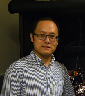
Jianing Yu
E-mail: jianing.yu(at)pku(dot)edu(dot)cn
Office: Lui Che Woo Building, Peking University
Lab: Lui Che Woo Building, Peking University
Research Area:
The brain uses efference copy signals to predict the sensory consequences of actions. This explains the observation that one cannot tickle themselves. However, in many cases, we still do not know the neural basis of efference copy signals, how they are linked to movement command, and how they interact with the sensory systems.
Our lab uses self-touch as a model to understand the neural basis of efference copy. Self-touch has been studied extensively in the human subject, where functional brain mapping and MEG recordings were applied to reveal the differential representations of self- versus external-touch in various brain regions. To understand at the circuit level how sensory signals from self-touch are processed, our lab develops novel tactile behaviors in head-fixed rodents and employs single-cell and population recordings to reveal differential representations of self-touch and external touch in distributed brain circuits, including the thalamus, cortex, and the cerebellum. We combine a wide range of techniques, including in vivo whole-cell recordings, brain slice electrophysiology, multi-channel single-unit recordings, intrinsic signal imaging, voltage and calcium imaging, optogenetics, chemogenetic and pharmacological manipulations. Together, we are interested in elucidating how animals track their own actions, as well as understanding multiple neurological diseases (including schizophrenia) that disrupt this basic function of the brain.
Another direction in the lab is to probe the neural circuits of the medial prefrontal cortex during executive functions. We will adopt a simple reaction time task for head-restrained rodents and test the animals’ cognitive ability in inhibitory control, for example, maintaining an action until a release cue is delivered. We want to understand the rules by which mPFC transmits information to downstream targets to inhibit or promote movement. Meanwhile, we will also investigate how long-range afferent inputs and local circuits give rise to diverse neural activity patterns in mPFC, focusing on biophysical and synaptic mechanisms.
Selected Publications:
1.Yu J, Hu H, Agmon A, Svoboda K. (2019) Recruitment of GABAergic Interneurons in the Barrel Cortex during Active Tactile Behavior. Neuron (in press). preprint, bioRix 554949.
2. Gutnisky D, Yu J, Hires SA, To MS, Bale MR, Svoboda K, Golomb D. (2017) Mechanisms underlying a thalamocortical transformation during active tactile sensation. PLoS Comput Biol. 7;13(6):e1005576.
3.Yu J*, Gutnisky D*, Hires SA, Svoboda K. (2016) Layer 4 fast-spiking interneurons filter thalamocortical signals during active somatosensation. Nature Neuroscience 19(12):1647-1657. (* equal contribution)
4. Hires SA, Gutnisky DA, Yu J, O'Connor DH, Svoboda K. (2015) Low-noise encoding of active touch by layer 4 in the somatosensory cortex. Elife 4:e06619.
5. Yu J, Ferster D. (2013) Functional coupling from simple to complex cells in the visually driven cortical circuits. Journal of Neuroscience 33(48):18855-66.
6. O'Connor DH, Hires SA, Guo ZV, Li N, Yu J, Sun QQ, Huber D, Svoboda K (2013) Neural coding during active somatosensation revealed using illusory touch. Nature Neuroscience 16(7):958-65.
7. Yu J, Ferster D (2010) Membrane potential synchrony in primary visual cortex during sensory stimulation. Neuron 68(6):1187-201.
8. Yu J, Daniels BA, Baldridge WH. (2009) Slow excitation of cultured rat retinal ganglion cells by activating group I metabotropic glutamate receptors. Journal of Neurophysiology 102(6):3728-39.
9. Yu J, Anderson CT, Kiritani T, Sheets PL, Wokosin DL, Wood L, Shepherd GM. (2008) Local-Circuit Phenotypes of Layer 5 Neurons in Motor-Frontal Cortex of YFP-H Mice. Frontiers in Neural Circuits 2008;2:6.
10. Weiler N, Wood L, Yu J, Solla SA, Shepherd GM. (2008) Top-down laminar organization of the excitatory network in motor cortex. Nature Neuroscience 11(3):360-6.
11. Hartwick AT, Bramley JR, Yu J, Stevens KT, Allen CN, Baldridge WH, Sollars PJ, Pickard GE. (2007) Light-evoked calcium responses of isolated melanopsin-expressing retinal ganglion cells. Journal of Neuroscience 27(49):13468-80.


